Histochem
Immunohistochemistry techniques have been used since the 1940s, when AH Coons and colleagues published their landmark paper describing the identification of tissue antigens using a direct fluorescence protocol (Coons AH, Creech HJ, Jones RN (1941) Immunological properties of an antibody containing a fluorescent group. Proc Soc Exp Biol Med 47:200–202).
Immunohistochemistry enables the visualization (using light or confocal microscopy) of the tissue distribution of specific antigens (or epitopes). The process localizes protein targets of interest by applying specific monoclonal or polyclonal antibodies to tissue surfaces in a process called antibody incubation. In recent years, the protocol has been refined with the advent of avidin-biotin immunohistochemistry.
This procedure makes use of the high affinity that avidin (or streptavidin) has for biotin. Multiple binding sites on the molecules result in the amplification of the tissue signal which is useful when trying to detect low copy number antigens.
A collection of useful immunohistochemistry protocols and other methods are available in the protocols section of the website. Protocol optimization is crucial to obtaining successful and reproducible results; the troubleshooting section has some useful suggestions for improving immunohistochemistry results.
_______
Histochem.net is an immunohistochemistry information resource for researchers. Please follow the all safety and precaution recommendations supplied with the products or equipment you use as well as your institutional guidelines. The protocols and suggestions on this website are not a substitute for formal training.
Exercise caution. Please follow the manufacturer’s recommendations and instructions when working with any reagent or system. The procedures posted on this website are for informational purposes only.
Avidin - Biotin & dab immunohistochemistry | |
| 1. Deparaffinize sections 2. Rehydrate In steps through to distilled water (dH2O). 3. Immerse sections in 0.5% v/v hydrogen peroxide/methanol for 10 minutes. 4. The antigen retrieval protocol can be carried out at this time, if this procedure is deemed necessary. 5. Wash the tissue sections in two changes of dH2O (2 x 5 minutes). 6. Immerse the sample slides in 50mM Phosphate Buffered Saline (PBS) 2 x 5 minutes. 7. Cover sections with blocking solution. 8. Remove the blocking solution. 9. Quickly circle the tissue section with a PAP pen (the tissue section should not be allowed to dry). 10. Add primary antiserum diluted in blocking solution and incubate overnight at 4ºC (or 60 minutes at 25ºC). 11. Wash in PBS buffer for 3 x 5 minutes. 12. Remove excess PBS buffer and incubate sections with biotinylated secondary antibody diluted in blocking reagent; incubate for 30 minutes at 25ºC. 13. Wash in PBS buffer for 3 x 5 minutes. 14. Remove excess PBS buffer and incubate sections with ABComplex/HRP for 30 minutes at 25ºC. 15. Wash in PBS buffer for 3 x 5 minutes. 16. Develop with 3 3’ diaminobenzidine tetrahydrochloride (DAB) 17. Rinse slides in gently running tap water. 18. Counterstain with Haematoxylin. 19. Dehydrate, clear and mount sections. |
Cryosectioning
1. Cool the cryostat to approximately -20 C (if it is too cold, the tissue will be difficult to cut)
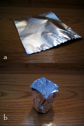
2. create a small bowl shaped aluminum foil basket to hold the tissue sample (see images)
3. pour some cryomatrix embedding agent into the foil and place the tissue sample into the liquid matrix
4. freeze the sample by dipping the foil into liquid nitrogen (use forceps to hold the foil)
5. place the frozen tissue/ cryomatrix block on the 'chuck' that guides the sample against the cryostat blade (adhere with a dab of cryomatrix solution
6. once the tissue block has frozen into place, move the block into place and cut the sections to the desired thickness (7 - 20 microns)
7. pick each slice up by pressing a glass microscope slide on the tissue so that it melts/adheres
8. leave each microscope slide containing tissue section in the cryostat chamber in a microscope rack to keep them frozen while you cut the rest of the block
9. wrap the rack in aluminium foil and store at -80 C
Sample Fixation
There are many methods of tissue sample fixation: from picric acid to bouins solution to paraformaldehyde. The following fixation protocol describes the use of 4% paraformaldehyde (PFA).
1. dissolve 4 g PFA in 50ml dH2O
2. add 2 drops 10N NaOH
3. add a stir magnet; stir and heat to 60 C - until solution is clear
4. add 50ml 2x PBS
5. adjust pH to 7.3
6. if working with cryosections, immerse slides with non-fixed tissue sections into PFA for 45 minutes
7. if preserving a whole tissue sample, immerse sample into PFA solution (the solution should be approximately ten times the volume of the tissue sample) overnight
Antigen Retrieval
Antigen retrieval (or antigen recovery) is performed to expose or retrieve antigens which have become masked by the tissue fixation process.
There are various protocols available. Some make use of heat induced epitope retrieval (HIER) while others apply enzymes.
The following antigen recovery protocol is microwave based. Studies have shown microwave antigen retrieval to be effective in unmasking epitopes in formalin fixed, paraffin wax embedded tissues.
1. Place slides in a plastic Coplin jar
2. Add citrate buffer - cover slides
3. Cover with a perforated film
4. Place Coplin jar into microwave
5. Microwave at medium power for 5 minutes
6. Add citrate buffer to replace evaporative losses
7. Microwave at medium for up to 5 minutes
8. Add more citrate buffer
9 . Allow slides to cool
10. Proceed with immunohistochemistry protocol
Slide Mounting
Mounting microscope slides refers to the coverslip mounting procedure. There are a number of different methods for mounting tissue sections. Glycerol can be used for temporary mounting. If a more permanent seal is important, Permount is recommended. Permount will not discolor with age.
1. dehydrate tissue sections through graded alcohol series and toluene (xylene)
2. remove the slide from toluene
3. place the slide horizontaly - tissue section facing upwards
4. apply 1-2 drops of permount onto the tissue section
5. gently place the coverslip onto the tissue sample
6. using forceps, apply slight pressure on the coverslip - eliminate any air bubbles
7. using tissue paper, blot any excess permount/toluene from around the edges of the coverslip
8. keep the slides flat; allow overnight drying (a slide oven at 75 C can be used as well)
9. although permount is permanent, coverslips can be removed by soaking slides in toluene
Periodic Acid Schiff
1. Deparaffinize the sections and rehydrate through graded alcohol steps
2. Oxidize in periodic acid solution for 10 minutes
3. Rinse in 4 changes of dH2O
4. Place in Schiff solution for 15 minutes
5. Rinse in 3 changes of dH2O
6. Immerse in 0.3% sodium borate for 15 seconds
7. Rinse in 4 changes of dH2O
8. Stain with hematoxylin (xx time)
9. Rinse in 3 changes of dH2O
10. Immerse in 0.3% sodium borate for 5 seconds
11. Rinse in 4 changes of dH2O
12. Dehydrate through graded alcohols
13. Clear in 3 changes of Xylene
14. Mount with synthetic resin
Useful options
Beautiful snippets
Amazing pages
Outstanding images
Immhunohistochemistry Terms and Definitions
Antigen
antigens are typically proteins, however, they can be any substance that triggers an immune response and antibody production
antigen retrieval is a protocol to expose antigens masked by fixation (see antigen retrieval protocol)
Avidin - biotin complex
there is a high affinity between avidin and biotin; immunohistochemistry makes use of this high affinity by using biotinylated antibodies to amplify tissue antigen signals
Counterstain
after immunohistochemical processing, counterstains are often applied to better visualize the results
Cryosection
cryosectioning is a tissue sectioning technique that cuts frozen tissue using a cryostat (a microtome operating at approximately -25 C)
Dewax
the process of removing paraffin from paraffin embedded tissues
Fixative
a solution that helps preserve tissue and cell morphology
IgG
immunoglobulin G - an antibody type normally present in the blood
IgM
immunoglobulin M - one of the 5 classes of immunoglobulins
Primary antibody
primary antibodies are glycoproteins that specifically bind antigens (epitopes)
Secondary antibody
a secondary antibody is raised to the Ig of the species from which comes the primary antibody; the secondary antibody will label the primary antibody
Triton
triton (triton x-100) is a nonionic surfactant; it is used to permeabilize cell memnranes to allow greater access to antibodies